Are you looking for an advanced imaging method for dental problems? Cone Beam CT (CBCT) is the solution. This technology provides detailed 3D images of teeth, jawbones, and related structures, strongly supporting diagnosis and treatment planning.
CBCT stands for Cone Beam Computed Tomography, a medical technique that uses specialized medical equipment – a computed tomography system – to create 3D images of the maxillofacial region. Unlike traditional 2D X-rays which only provide flat images, CBCT allows doctors to observe anatomical structures from all angles, helping to accurately detect potential problems.
What is Cone Beam CT imaging?
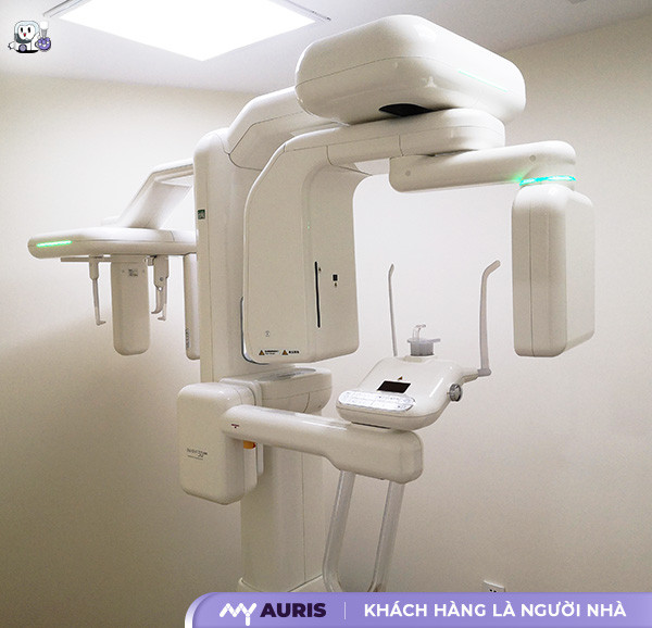
Cone beam CT imaging (cone beam computed tomography technique) is an advanced imaging method in modern dentistry. This technique uses special technology to reconstruct sharp 3D images of soft tissues and dental tissues with just a single scan. During the scan, the device rotates 360 degrees around the patient’s head, capturing every view of the teeth and the area to be examined. From there, the machine produces high-resolution images, assisting doctors in developing detailed and precise treatment plans.
Compared to traditional dental X-rays, cone beam CT imaging offers many outstanding advantages:
Provides clearer and sharper 3D images by focusing on the area of interest.
Obtains multiple different views, including hard-to-observe areas, with just a single scan.
It is a non-invasive, painless, and absolutely safe method for patients.
The equipment has high durability, ensuring long-lasting and stable image quality.
Thanks to these advantages, cone beam CT imaging is widely applied in many modern dental services, especially:
Dental Implant Placement: The images obtained help dentists assess bone density and width, thereby determining whether bone grafting is needed and selecting the appropriate implant post.
Endodontic Treatment: Helps precisely locate impacted teeth, root canals, and internal structures for effective and safe treatment.
Orthodontics: Provides detailed data on tooth-jaw structure for precise braces planning, optimizing aesthetic and functional results.
Wisdom Tooth Minor Surgery: Allows evaluation of the relationship between the wisdom tooth root and nerves, assisting dentists in precise extraction point localization, avoiding damage to surrounding structures.
Is Cone Beam CT imaging safe?
Cone Beam CT (CBCT) imaging is a modern diagnostic imaging method that uses X-rays to reconstruct detailed 3D images of the jawbones, teeth, and related structures. However, many patients still wonder about the level of radiation safety when undergoing this technique. Here is important information to help you feel more at ease:
Low Radiation Dose: Compared to traditional X-ray techniques, CBCT uses a significantly lower radiation dose. Specifically, according to data from Planmeca machines, this advanced technology can reduce the radiation dose by up to 77%, much lower than conventional nano CT, helping to minimize X-ray impact on the body.
Non-Invasive Technique: Cone Beam CT imaging is a non-invasive technique that does not require touching soft tissues or performing invasive procedures. Throughout the process, the patient only needs to sit or lie still, completely pain-free and absolutely safe.
Short Scan Time: A CBCT scan only takes a few minutes, thereby minimizing X-ray exposure time and providing comfort to the patient, especially suitable for the elderly and children.
CBCT is currently an indispensable diagnostic tool in many dental applications such as: dental implant placement, orthodontics, and maxillofacial surgery. The accuracy and safety of the images help doctors develop optimal and highly effective treatment plans.
Although it is a safe method, patients should still note some special cases:
Pregnant Women: It is necessary to consult a doctor because, even with a low radiation dose, any risk to the fetus should be avoided.
Contrast Medium Allergy: If you have had a reaction before, you need to inform your doctor clearly before the procedure.
Special Health Conditions: Full medical information must be provided for appropriate consultation.
To ensure effectiveness and radiation safety, patients should choose a reputable dental facility with modern equipment, using new technologies like Planmeca, along with an experienced team of doctors in diagnosis and treatment.
Applications of Cone Beam CT in Dentistry
CBCT uses a cone-shaped X-ray beam to obtain 3D images in a single scan, with high spatial resolution and a radiation dose typically lower than traditional CT:
How is CBCT applied in Pre-Surgical Diagnosis?
Maxillofacial surgical planning requires high precision. CBCT provides detailed 3D images of the jawbones, temporomandibular joint, maxillary sinus, and adjacent structures. This allows imaging specialists to accurately assess the location, size, and shape of tumors, bone lesions, evaluate bone density, and survey the paths of nerves and blood vessels. Consequently, surgeons can create detailed surgical plans, minimize risks, and enhance treatment effectiveness.
Applications of CBCT in Dental Implants
Dental implants are an effective method of tooth restoration. CBCT plays a crucial role in evaluating jawbone volume and quality, determining the optimal implant placement. Accurate 3D images help dentists select the appropriate implant size and type, ensuring successful and long-lasting implant procedures. CBCT also helps detect abnormalities in the jawbone, maxillary sinus, and nerves, thereby enabling dentists to make suitable treatment decisions and avoid potential complications. The use of CBCT in implant placement increases the success rate and minimizes risks.
Applications of CBCT in Sinus Assessment and Orthodontics
CBCT provides detailed images of the maxillary sinus, aiding in the diagnosis of sinus pathologies such as sinusitis and sinus polyps. In orthodontics, CBCT supports the evaluation of jawbone, tooth, and soft tissue structures, helping doctors create effective orthodontic treatment plans. 3D images help determine the position and eruption direction of teeth, assess the degree of crowding, and thus select the appropriate orthodontic method. The use of CBCT in orthodontics helps shorten treatment time and achieve optimal aesthetic results.
Applications of CBCT in Endodontics
In endodontics, CBCT helps diagnose periapical lesions, evaluate the shape and number of root canals, and detect accessory canals. This information helps endodontists perform more effective root canal treatments and reduce the risk of reinfection. CBCT also assists in evaluating cases of fractured instruments within root canals, helping doctors determine appropriate management plans.
How Does Cone Beam CT Work?
The workflow of a CBCT scanner is summarized as follows:
Patient Positioning: The patient sits or stands still in the CBCT scanner. Correct positioning is crucial to ensure accurate and clear images. The tomography system is designed to minimize patient discomfort.
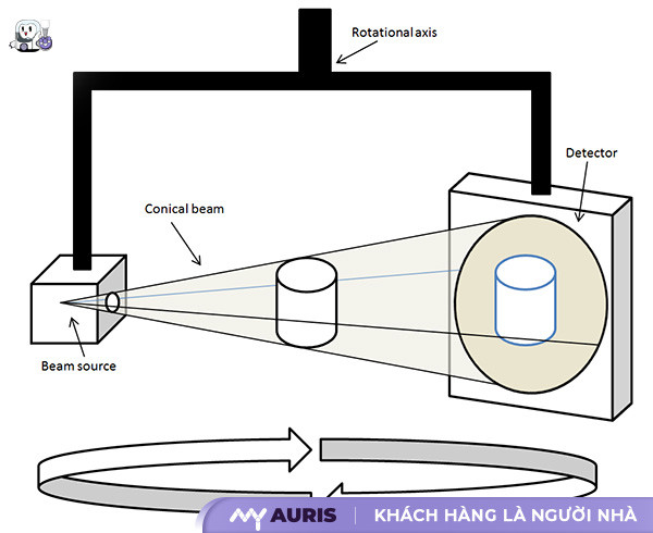
X-ray Source and Detector Rotate Around the Patient: A CBCT scanner uses a cone-shaped X-ray source and a detector that rotates around the patient. During the scan, the X-ray source emits a cone-shaped X-ray beam that passes through the area of interest, such as the jawbone.
Detector Records Data: The detector records the intensity of the X-rays after they pass through the patient’s body. This data is converted into electronic signals.
Data Processed by Software: The electronic signals from the detector are transmitted to a computer and processed by specialized diagnostic software. This software uses complex algorithms to reconstruct 3D images of the scanned area.
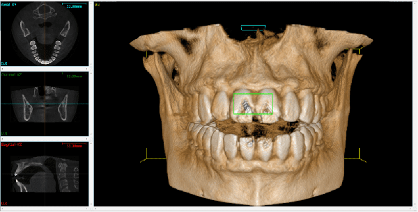
3D Images Displayed: The software generates 3D images that can be viewed on a computer screen. Imaging specialists or dentists can examine these images in various cross-sectional planes, zoom in, zoom out, rotate, and analyze them to diagnose pathologies. X-ray technicians assist during the imaging and image processing steps.
Superior Advantages of Cone Beam CT Compared to Traditional X-rays in Dentistry
Unlike 2D X-rays that only provide planar images, CBCT generates 3D images of teeth, jawbones, maxillary sinuses, and the temporomandibular joint. This allows imaging specialists to observe anatomical structures from all angles, detecting abnormalities that 2D X-rays would struggle to identify.
Although it is a medical technique that uses radiation, the radiation dose of CBCT is usually significantly lower than that of traditional computed tomography (CT) scans.
CBCT scan time is extremely fast, often under 5 seconds, which minimizes the patient’s exposure to radiation. The scanning process is also very simple, causing no discomfort or pain. Patients only need to sit still for a few seconds, and the CBCT scanner will automatically collect data and create 3D images. This advantage is particularly useful for sensitive patients, children, or those with claustrophobia.
When is Cone Beam CT imaging needed?
Cases where CBCT imaging should be considered:
Dental Implant Placement: Accurately determining jawbone volume and the position of critical anatomical structures like nerves and maxillary sinuses is a key factor for successful implant placement. 3D images from CBCT provide detailed information, helping doctors optimize implant planning and minimize risks. This diagnostic method is superior to traditional 2D X-rays.
Maxillofacial Surgery: For complex surgeries such as impacted wisdom tooth extraction, jawbone cysts, and congenital maxillofacial deformities, CBCT helps doctors clearly visualize bone structures, blood vessels, and nerves. This allows for precise surgical planning, minimizing complications, and enhancing treatment effectiveness.
Orthodontics: CBCT supports diagnosis and orthodontic treatment planning, especially in difficult cases such as misaligned teeth or complex malocclusions. 3D images help precisely determine tooth and jawbone positions, assisting doctors in creating appropriate treatment protocols.
Sinus and Temporomandibular Joint Assessment: CBCT allows for detailed evaluation of maxillary sinus and temporomandibular joint structures, detecting abnormalities such as sinusitis and joint degeneration. This information helps doctors make accurate diagnoses and effective treatment methods.
Endodontics: In some complex endodontic cases, CBCT helps accurately determine the position and shape of root canals, supporting more effective treatment.
Best Cone Beam CT Scanners Today
Below are some leading Planmeca machine lines, known for their image quality, high spatial resolution, and low radiation dose:
Planmeca ProMax® 3D Mid
ProMax 3D Mid is an ideal medical device for most clinical applications in dentistry. This CBCT scanner provides high-quality 3D images with various scanning volumes, meeting diagnostic needs from dental implants to maxillofacial surgery. The fast scanning time reduces patient waiting time. Integrated diagnostic software supports doctors in easily analyzing images.
Planmeca ProMax® 3D Plus
ProMax 3D Plus is aimed at large clinics and hospitals. This computed tomography system features advanced technology, allowing for large volume scans with superior spatial resolution. The applications of CBCT in dentistry are expanded, including dental material research. Its ability to integrate with surgical navigation systems enhances accuracy in complex cases.
Planmeca ProMax® 3D Classic
ProMax 3D Classic is an economical choice that still ensures good image quality. This 3D scanner in dentistry is suitable for small-scale clinics, focusing on basic diagnostic applications such as implants and orthodontics. Integrated dental treatment planning software helps optimize workflows.
Planmeca ProMax® 3D s
ProMax 3D s focuses on maxillofacial imaging, providing detailed images of teeth, maxillary sinuses, and the temporomandibular joint. This CT scanner is useful for diagnosing bite problems, temporomandibular disorders, and maxillofacial pathologies. The low radiation dose ensures patient safety.
Planmeca Viso™ G7
Viso G7 represents a new step forward in dental imaging. This CBCT scanner is equipped with advanced technology, allowing for large volume scans with extremely high spatial resolution. The ultra-fast scan time of under 5 seconds enhances the patient experience. Its automatic analysis and reporting capabilities effectively assist imaging specialists in diagnosis.
Planmeca Viso™ G5
Viso G5 delivers high-quality 3D images at a reasonable cost. This CBCT scanner uses a cone-shaped X-ray beam, obtaining 3D images in a single scan. Its high spatial resolution and typically lower radiation dose compared to traditional CT are particularly useful in dental and maxillofacial diagnosis.